Vinayaka Missions Medical College is situated in the picturesque views of Bay of Bengal. This institution was established in the year 1996 by our Former Chairman, (Late) Dr.A.Shanmugasundaram, a noble Philanthropist and Educationist of extraordinary vision in the field of education who is the founder of Vinayaka Missions University belonging to the Thirumuruga Kirupananda Variyar Thavathiru Sundara Swamigal Medical Educational & Charitable Trust.
Anatomy is a branch of medicine that studies the structure of the Human body in Macroscopic & Microscopic and Developmental aspects. This branch specializes in various research aspect specifically genetic research, Cadaveric dissection, Histological study & Tissue processing. Department of Anatomy generates significant contributions to Mission of the College. Department currently has well trained post graduate qualified faculty members with supporting staff.
The broad goal of the teaching of undergraduate students in anatomy aims at providing comprehensive knowledge of the gross and microscopic structure and development of human body to provide a basis for understanding the clinical correlation of organs or structures involved and the anatomical basis for the disease presentations.
To achieve highest standard of imparting knowledge of Anatomy.
To prepare and develop the department for Post graduation level.
To lay a strong foundation for medical education with emphasis on clinically relevant course material, so as to address a crucial need for patient care & support and medical research in basic sciences.
Comprehend the normal disposition, clinically relevant inter-relationships, functional and cross sectional anatomy of the various structures in the body.
Indentify the microscopic structure and correlate elementary ultra structure of various organs and tissues and correlate the structure with the functions as a prerequisite for understanding the altered state in various disease processes.
Comprehend the basic structure and connections of the central nervous system to analyze the integrative and regulative functions of the organs and systems. He / She shall be able to locate the site of gross lesions according to the deficits encountered.
Demonstrate knowledge of the basic principles and sequential development of the organs and systems; recognize the critical stage of development and the effects of common teratogens. He / She shall be abnormalities.
At the end of the course the student shall be able to: Indentify and locate all the structures of the body and mark the topography of the living anatomy.
Identify the organs and tissues under the microscope: Understand the principles of karyotyping and identify the gross congenital anomalies; Understand the principles of newer imaging techniques like Ultra sound, Computerized Tomography Scan (CTS); Interpretation of Plain and contrast X-rays.
Understand clinical basis of some common clinical procedures i.e. intramuscular and intravenous injection, lumbar puncture, kidney biopsy etc.
The Anatomy department facilitates all the learning aspects of the Morphological science in the first year of curriculum. Well qualified faculty members are teaching the Gross Anatomy, Microscopic Anatomy or Histology, Developmental Anatomy, Genetics, Radiology surface marking and Osteology under the regulation of VMRF University.
The Anatomy department trains the students by using both traditional & innovative teaching methods. The Anatomy department actively takes part in adopting newer teaching methods like small group discussion, problem based learning, demonstration and student seminar and integrated teaching. The traditional lecture classes enhanced by the advance technologies like computer aided smart board and audio visual aids.
The Anatomy department trains Under Graduate Medical Students, Basic science classes for Post Graduate students, Students of FAHS and Nursing students.
VMRF University offers PhD programme research in Anatomy and every year 3 to 5 candidates are register their PhD in Anatomy.
Periodic formative assessment tests conducted by the department of Anatomy. It includes three terminal examination and model exam. Code number used to be given for each assessment test to avoid bias. Summative assessment conducted by VMRF University at the end of the first year.
After the first terminal assessment poor performers & slow learners were identified & counselling given. For them special arrangements, to improve their performance, were also made.
The following teaching - learning methods were adopted
All faculty were assigned by Institute to do mentorship for five student. Apart form general mentor programme ,Small group (17) of students were given in a care of each staff member who analysis individual students, short coming and provided counseling regarding their performance of Anatomy.
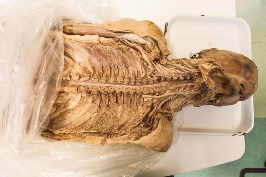
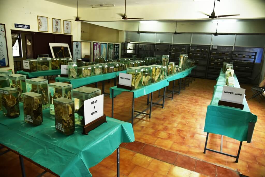
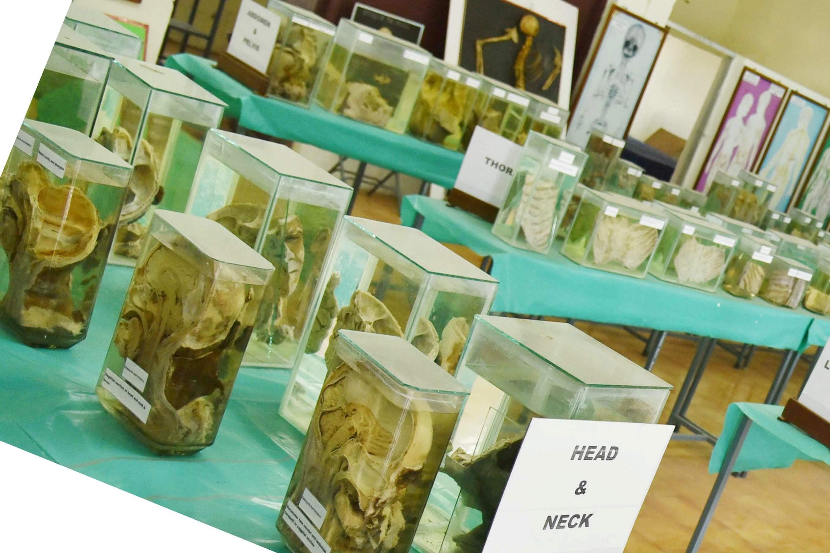
The excellent museum spread over 235 sq.mt., exhibits more than 100 dissected cadaveric and foetal specimens. The museum also displays sections of human body, radiographs, human skeletons, radiographs and developmental anatomy models. The museum collection is presented in several sections, e.g. Head and Neck, Thorax, Abdomen and Pelvis, Limbs, Sectional anatomy, Nervous system and Embryology. New specimens and models are continuously added to the collection. Presently, genuine efforts are being made to promote the museum as a lively source of interaction for health science students and also the public.
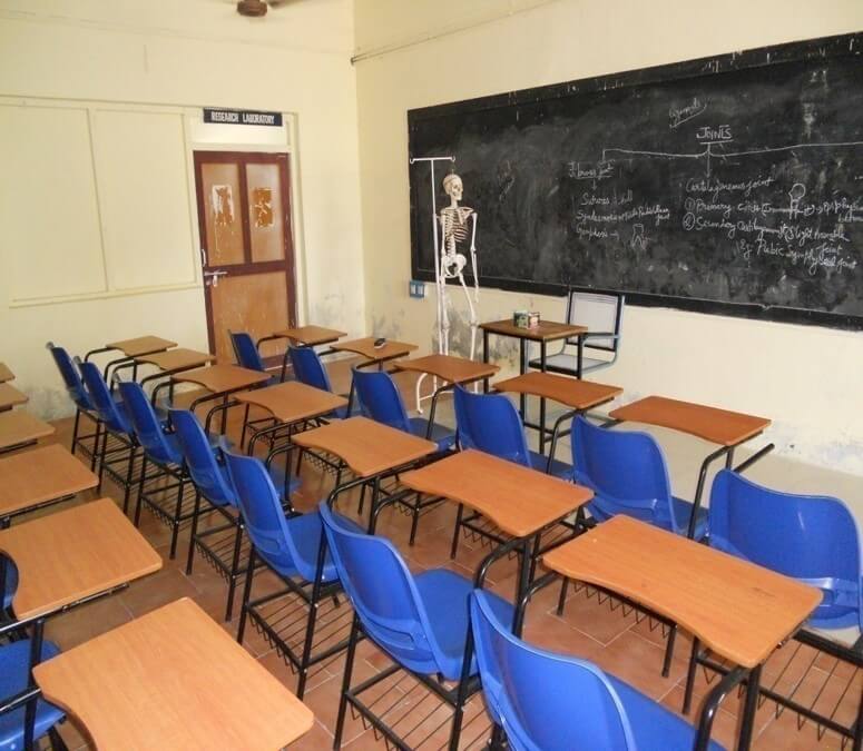
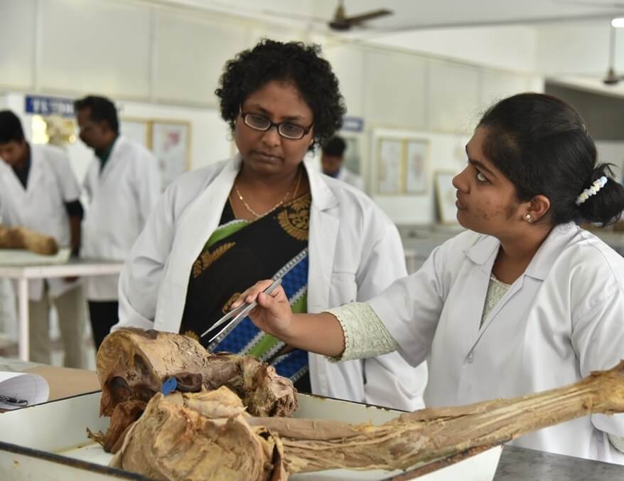
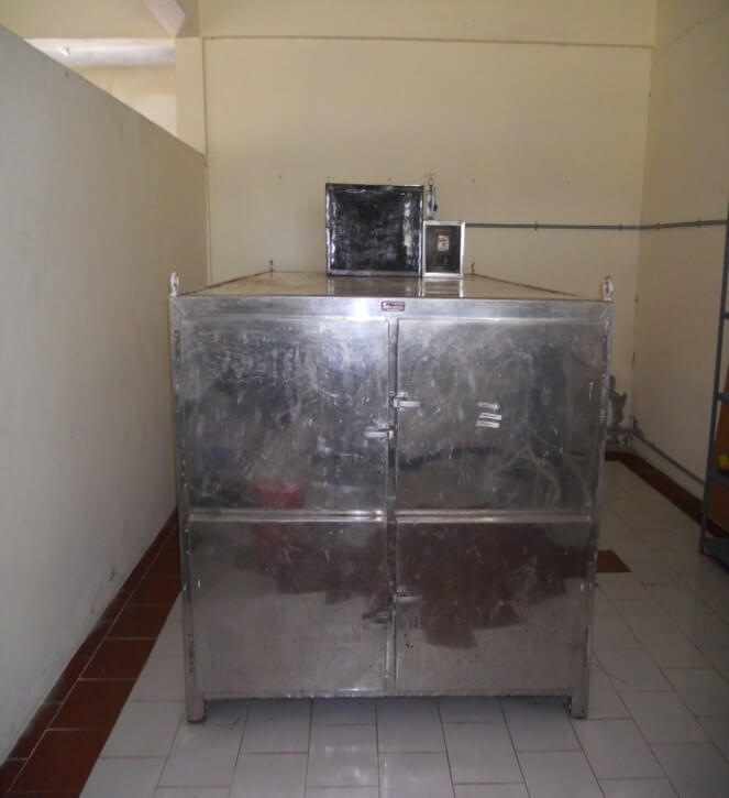
The 345 sq mt. clean and brightly-lit, hall ensures adequate space for more than 150students at a time. The excellent cadaver preservation facilities include cold cabinets which can accommodate 8 cadavers. Formalin tanks for cadaver preservation and wash area is also available. Moreover, well preserved specimens of all parts of the human body are easily available to the students. The teaching-learning process is enhanced by normal X-ray, MRI, CT scan images and embryology models. The individual bones and articulated skeletons are also made accessible to the students during working hours. The dissection hall is well equipped with anthropometric instruments, X-ray view boxes and instruments essential for preparation of anatomical specimens, e.g. band saw, circular saw, brain knife etc.
Two demonstration rooms, each of 60 sq. mts.,accommodating 75 students and equipped with appropriate audio visual aids, add to the teaching ambience of the department.
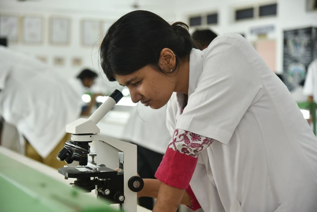
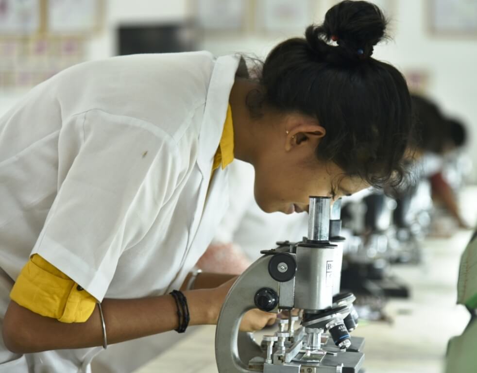
The department has a fully equipped histology unit spread over an area of 225 sq.mt. which can accommodate 90 students at a time. Students are provided with individual microscopes and slides during routine practical classes and a binocular microscope with CCTV attachment for the projection of slides for teaching. Colored charts of labelled microanatomy sections of all tissues are displayed for students for reference during the routine practical sessions. The lab is also well equipped with tissue processing instruments, e.g. paraffin embedding bath, incubator, microtome etc. to make slides for teaching purposes.
Bodies received from appropriate sources after due processes are embalmed in this 17 sq. mts. room equipped with two embalming machines before being transferred to the storage tanks.
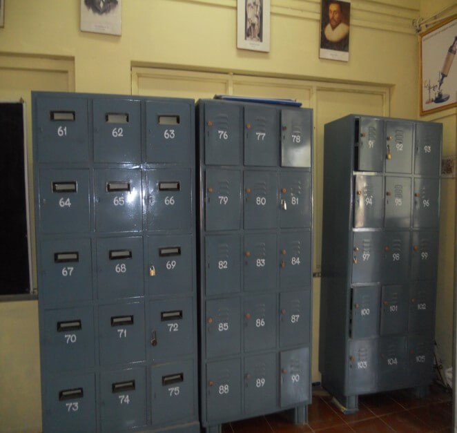
Three storage tanks of 3, 3 and 2 sq. mts. respectively are used for the storage of the embalmed bodies, namely, the cadavers. The number of cadavers will be proportionate to the number of students.150 lockers for storing student belongings during dissection hours are provided in the Anatomy Dissection Hall corridor. Research Lab is equipped with the instruments needed for the research activity in various fields of Anatomy like Gross anatomy , embryology, histology, genetics, etc..
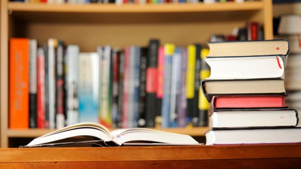
The departmental library consists of an excellent collection of books on life sciences. In addition to a series of basic and standard reference books in gross anatomy, histology, embryology, neuroanatomy, it also has vast collection of books from physiology, biochemistry, medicine, surgery, paediatrics, radiology, gynaecology etc. This facilitates the learning of clinical anatomy correlation with other clinical areas.
Topic | Researcher | Remarks |
---|---|---|
Morphological variation of pulmonary fissure, lobes and bronchial tree | Dr.Santhini Arulselvi | ongoing |
Morphological study of basilar artery and its variations | Mr.J.Kalaivannan | ongoing |
Anatomical significance of right conus artery in south Indian population | Mrs.T.Udaya Sankari | ongoing |
Role of nuclear anomalies and DNA damage in early diagnosis of cervical cancer | Dr. Sohinder Kaur | ongoing |